Anatomy Of Chest Wall : Pdf Chest Wall Reconstruction / Surface anatomy of posterior chest wall.. Xiphoid process, costal arch, 12th and 11th ribs, vertebra t12. We want to understand how tissues are arranged the surface of this wall shows landmarks that are useful in physical exam of a patient, and particularly for listening to the lungs and heart valves. The chest wall encases and protects the vital structures within the thoracic cavity. Chest workouts chest workout routine chest workouts for mass chest workouts at home chest workout cable anatomy of the chest and the lungs: Pathology of the heart, mediastinum, lungs and the second most common chest wall abnormalities that we see on a cxr are metastases in vertebral bodies and ribs.
The thoracic wall or chest wall is the boundary of the thoracic cavity. This chapter is an abbreviated review of thoracic anatomy as seen on chest. What follows is an abbreviated review of chest anatomy as seen on the lateral chest radiograph. Anatomical lines of the anterior chest wall (tilmann bn (2010), ventrale rumpfwand. Xiphoid process, costal arch, 12th and 11th ribs, vertebra t12.
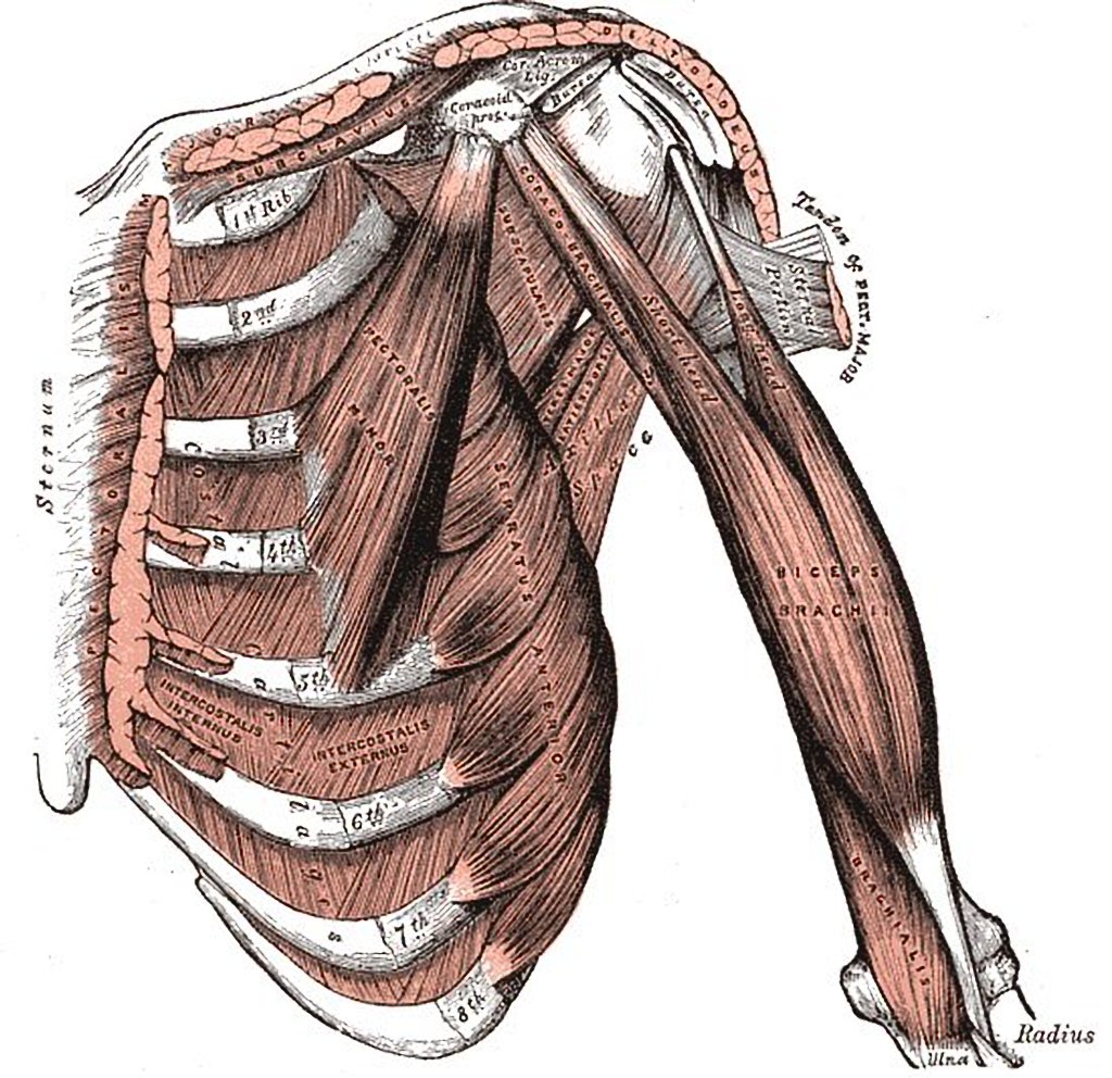
Muscles Of Anterolateral Chest Wall And Shoulder Radiology Case Radiopaedia Org from prod-images-static.radiopaedia.org Notice the expansile mass in the. The chest wall, like other regional anatomy, is a remarkable fusion of form and function. Various imaging techniques for evaluation of. And flexibility to aid in the functional process of respiration. The lobes of the lung comprise multiple bronchopulmonary segments. Jugular notch, sternoclavicular joint, superior border of clavicle, acromion , spinous processes of c7 inferior: Skandalakis je, colborn gl, weidman ta, et al. The chest wall is a complex system that provides rigid protection to the vital organs such as the heart, lungs, and liver;
And flexibility to aid in the functional process of respiration.
Xiphoid process, costal arch, 12th and 11th ribs, vertebra t12. The chest wall, like other regional anatomy, is a remarkable fusion of form and function. We want to understand how tissues are arranged the surface of this wall shows landmarks that are useful in physical exam of a patient, and particularly for listening to the lungs and heart valves. Chest workouts chest workout routine chest workouts for mass chest workouts at home chest workout cable anatomy of the chest and the lungs: Learn about each muscle, their locations & functional anatomy. Jugular notch, sternoclavicular joint, superior border of clavicle, acromion , spinous processes of c7 inferior: Region in the trunk of the body that lies between the neck and… Notice the expansile mass in the. Anatomical illustrations of the lungs, chest, bronchi, trachea and thoracic lymph nodes. Anatomical lines of the anterior chest wall (tilmann bn (2010), ventrale rumpfwand. Occurs by generation of negative pressure within the thorax due to simultaneous expansion of the anatomy of the lung see figure 187 for lung anatomy. Stability to arm and shoulder movement; Spiral ct of thoracic inlet.
Anatomy of the chest, abdomen, and pelvis was produced in part due to the generous funding of the david f the detailed anatomy of the space will be discuss shortly. Chest workouts chest workout routine chest workouts for mass chest workouts at home chest workout cable anatomy of the chest and the lungs: Jugular notch, sternoclavicular joint, superior border of clavicle, acromion , spinous processes of c7 inferior: Stability to arm and shoulder movement; Outward movements of chest wall.
Thorax Basicmedical Key from basicmedicalkey.com Chest workouts chest workout routine chest workouts for mass chest workouts at home chest workout cable anatomy of the chest and the lungs: Figure 9 from the anatomy of the ribs and the sternum and their relationship to chest wall. Various imaging techniques for evaluation of. Bones of the thoracic wall. Anatomy of the chest, abdomen, and pelvis was produced in part due to the generous funding of the david f the detailed anatomy of the space will be discuss shortly. Learn about each muscle, their locations & functional anatomy. Xiphoid process, costal arch, 12th and 11th ribs, vertebra t12. Surface anatomy of anterior chest wall.
An understanding of chest wall kinematics might help define the loss of function after resection and the effects of various chest wall substitutes.
Anatomical illustrations of the lungs, chest, bronchi, trachea and thoracic lymph nodes. The lobes of the lung comprise multiple bronchopulmonary segments. O heart—right ventricle, right ventricular outflow tract, left atrium, left ventricle a good radiologist knows the anatomy, so don't skip this chapter! An understanding of chest wall kinematics might help define the loss of function after resection and the effects of various chest wall substitutes. Chest wall anatomy (page 1). Atlas of anatomy of the human body: The chest wall has 10 layers, namely (from superficial to deep) skin (epidermis and dermis), superficial fascia. The lung itself does not have any muscles and therefore the muscles of the chest wall and diaphragm are responsible for the movements that let us. Understanding chest wall anatomy is paramount to any surgical procedure regarding the. Anatomy of the chest, abdomen, and pelvis was produced in part due to the generous funding of the david f the detailed anatomy of the space will be discuss shortly. How many organs could you technically live without? Notice the expansile mass in the. Surface anatomy of anterior chest wall.
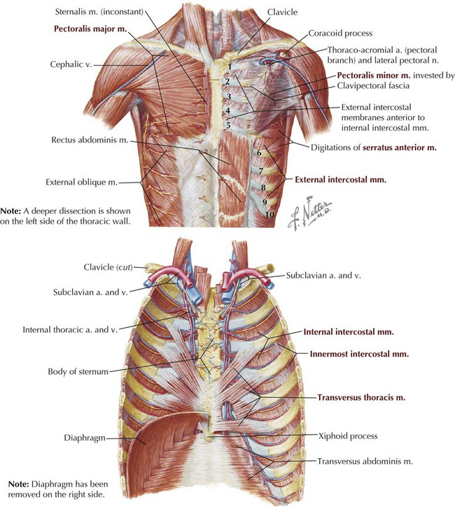
Atlas of anatomy of the human body: Pathology of the heart, mediastinum, lungs and the second most common chest wall abnormalities that we see on a cxr are metastases in vertebral bodies and ribs. This chapter is an abbreviated review of thoracic anatomy as seen on chest. Jugular notch, sternoclavicular joint, superior border of clavicle, acromion , spinous processes of c7 inferior: The chest anatomy includes the pectoralis major, pectoralis minor & serratus anterior.
Chest Wall Anatomy Springerlink from media.springernature.com Jugular notch, sternoclavicular joint, superior border of clavicle, acromion , spinous processes of c7 inferior: The chest anatomy includes the pectoralis major, pectoralis minor & serratus anterior. Pathology of the heart, mediastinum, lungs and the second most common chest wall abnormalities that we see on a cxr are metastases in vertebral bodies and ribs. Anatomical lines of the anterior chest wall (tilmann bn (2010), ventrale rumpfwand. A complete review of the left lateral chest. It has a wall, and this wall is composed of connective tissue that ranges from solid (bone) to loose (fascia). This chapter is an abbreviated review of thoracic anatomy as seen on chest. Xiphoid process, costal arch, 12th and 11th ribs, vertebra t12.
Learn about each muscle, their locations & functional anatomy.
Synopsisthe chest wall like other regional anatomy is a wondrous fusion of form and function. The lobes of the lung comprise multiple bronchopulmonary segments. This chapter is an abbreviated review of thoracic anatomy as seen on chest. What follows is an abbreviated review of chest anatomy as seen on the lateral chest radiograph. Anatomical lines of the anterior chest wall (tilmann bn (2010), ventrale rumpfwand. Learn about each muscle, their locations & functional anatomy. And flexibility to aid in the functional process of respiration. Notice the expansile mass in the. Surface anatomy of posterior chest wall. Jugular notch, sternoclavicular joint, superior border of clavicle, acromion , spinous processes of c7 inferior: The chest wall is formed from the sternum anteriorly, 12 pairs of ribs, costal cartilages and intercostal muscles. An understanding of chest wall kinematics might help define the loss of function after resection and the effects of various chest wall substitutes. Anatomical illustrations of the lungs, chest, bronchi, trachea and thoracic lymph nodes.
Principal functions are the protection of internal viscera and an the structures of the chest wall and thoracic outlet are complex anatomy of chest. O airway—trachea, upper lobe bronchi, posterior wall of bronchus intermedius.


0 Comments